A new MIT-led study maps the three-dimensional atomic architecture of relaxor ferroelectrics, offering a clearer path to designing better sensors, energy devices and imaging technologies.
For decades, relaxor ferroelectrics have helped power some of the most important sensing technologies in modern life, including medical ultrasound machines, microphones and sonar systems. Engineers knew these materials behaved in extraordinary ways. They could convert electrical signals into mechanical motion, respond strongly to external fields and support sensitive detection systems. What remained elusive was the inner structure that made those abilities possible.
Now, researchers from the Massachusetts Institute of Technology and collaborating institutions say they have directly mapped the three-dimensional atomic structure of a relaxor ferroelectric for the first time, resolving a long-standing mystery in materials science. The findings, reported in the journal Science and highlighted by ScienceDaily and MIT News, give scientists a more detailed picture of how electric charges and chemical disorder are arranged inside the material at the nanoscale.
The work matters because relaxor ferroelectrics are not obscure laboratory curiosities. They are functional materials used in devices that convert energy between electrical and mechanical forms. In medical ultrasound, related piezoelectric materials help generate and detect sound waves that pass through the body, producing images used in prenatal care, cardiology, cancer diagnosis and emergency medicine. In sonar and sensing systems, similar properties allow devices to transmit or detect signals with high sensitivity.
Yet the materials have long presented a paradox. Their performance depends on disorder, but that disorder has been difficult to observe directly. Traditional models could describe some average behavior, but they could not fully reveal how atoms and charges are arranged in three dimensions. That left researchers with useful materials but incomplete explanations.
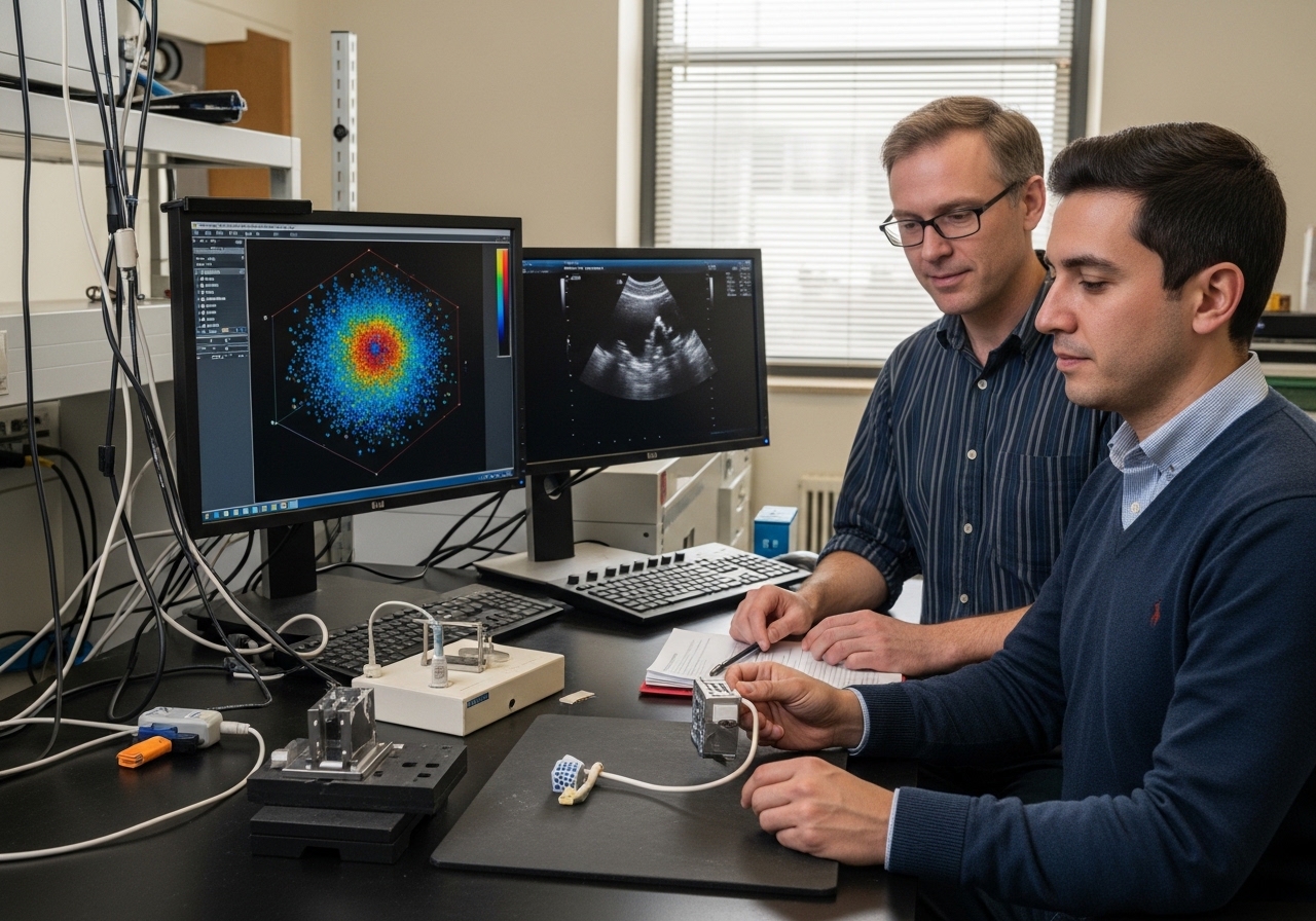
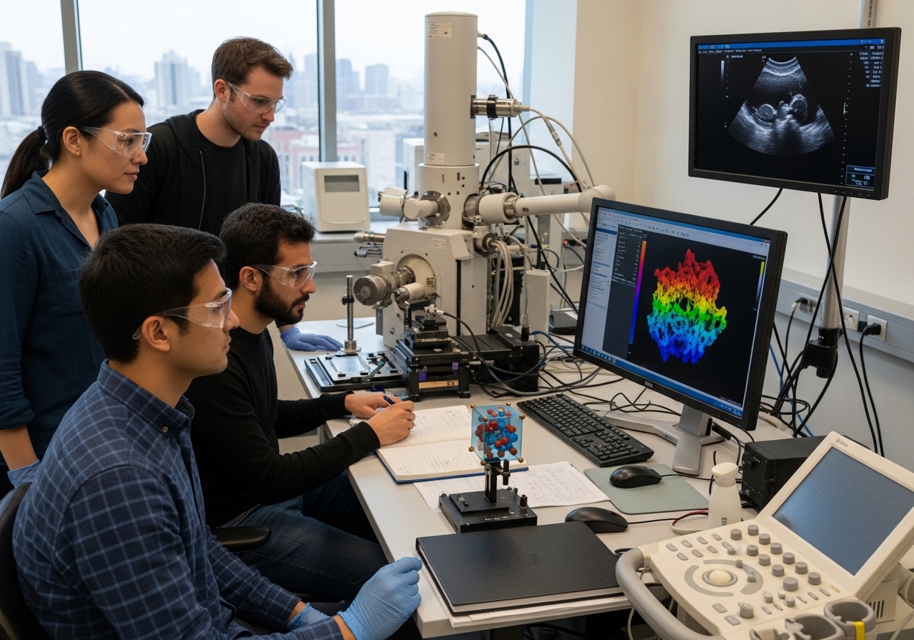
The MIT-led team focused on a lead magnesium niobate-lead titanate alloy, a type of relaxor ferroelectric used in sensors, actuators and defense-related systems. To examine it, the researchers used a technique called multi-slice electron ptychography, or MEP. The method scans a nanoscale beam of high-energy electrons across a sample and records diffraction patterns. Because neighboring scan positions overlap, computational algorithms can reconstruct three-dimensional information about the material’s atomic structure and electron wave function.
The result was a more detailed map than previous techniques had been able to provide. The researchers found a hierarchy of chemical and polar structures extending from individual atoms to larger mesoscopic features. They also discovered that regions with different polarization were significantly smaller than leading simulations had predicted. In simple terms, the material’s internal landscape was more intricate and more locally varied than scientists had assumed.
That discovery matters for the way researchers design new materials. Computer models are now central to materials science, especially as artificial intelligence and advanced simulations become more common. But a model is only useful if it reflects reality. James LeBeau, MIT’s Kyocera Professor of Materials Science and Engineering and corresponding author of the study, said the work gives researchers a better foundation for predicting and engineering desired material properties. Without accurate structural information, scientists risk building models that reproduce assumptions rather than physical truth.
The study also showed that chemical disorder had not been fully captured in earlier models. Co-first authors Michael Xu and Menglin Zhu, both MIT postdoctoral researchers, said the team was able to merge experimental observations with simulations to refine those models. That link between measurement and theory is one of the study’s most important contributions. It does not merely produce an image; it provides a way to test whether the theoretical picture of the material is correct.
Relaxor ferroelectrics are especially valuable because of the way their atoms shift and generate electric polarization. In a conventional ferroelectric, regions of polarization often behave in more orderly ways. In relaxor ferroelectrics, local variations in chemistry and charge create complex nanoregions that respond strongly to external electric fields. Those interactions help explain why the materials can be so useful in energy storage, sensing and electromechanical applications.
The new research suggests that the internal polar regions are not randomly arranged in the simple way some models had assumed. Instead, the correlations between those regions, and the influence of different chemical species, appear to be central to the material’s behavior. By showing how individual atomic species affect polarization depending on charge state, the study gives researchers a more precise framework for understanding the material’s response.
For medical technology, the implications are indirect but important. The study does not immediately produce a new ultrasound probe or diagnostic device. It does, however, improve the scientific foundation behind materials that can be used in transducers, sensors and imaging systems. Better models could eventually help engineers design materials with stronger sensitivity, greater stability, broader bandwidth or more efficient energy conversion.
That could matter in future ultrasound systems, where improvements in resolution, signal strength and miniaturization are highly valuable. Ultrasound is widely used because it is noninvasive, portable and comparatively affordable, but its performance still depends on the quality of the materials that generate and detect acoustic signals. If scientists can better control the nanoscale structure of relaxor ferroelectrics, they may be able to tune materials for more demanding imaging applications.
The findings also extend beyond medicine. MIT News noted that the work could help refine materials for next-generation computing systems, energy devices and advanced sensors. Relaxor ferroelectrics are of interest for memory storage, actuation, energy harvesting and other technologies where strong electromechanical or dielectric responses are useful. A clearer structural map could help researchers identify which forms of disorder improve performance and which limit it.
The study also highlights the rising power of electron ptychography in materials research. Complex materials often resist easy classification because their properties emerge from disorder, defects or local variation rather than from perfect repeating crystal structures. Traditional crystallography is excellent at describing average order, but many advanced materials work precisely because they are not perfectly ordered. Techniques such as MEP allow scientists to probe that complexity directly.
That is a major shift. For years, researchers had to infer the nanoscale behavior of relaxor ferroelectrics from indirect measurements and simulations. The new work provides direct volumetric evidence, linking three-dimensional polar structure with molecular dynamics calculations. According to the researchers, this is the first time such a connection has been made in an electron microscope for relaxor ferroelectrics.
The broader lesson is that some of the most useful materials in modern technology may still contain hidden structures that science has not fully seen. Engineers can build devices from them, manufacturers can scale them, and doctors can rely on equipment that uses them, even while the deepest atomic explanation remains incomplete. The MIT-led study narrows that gap between practical use and fundamental understanding.
The next step will be using this knowledge to guide design. Researchers will need to test whether the improved models can predict new compositions or processing methods that enhance performance. They will also need to determine how broadly the findings apply across the larger family of relaxor ferroelectrics, including compositions used in commercial and experimental devices.
For now, the discovery provides something materials scientists have sought for decades: a clearer view inside a class of materials whose usefulness exceeded their explanation. In a field where tiny atomic shifts can determine the performance of medical imaging equipment, sensors and future electronics, seeing the hidden structure is more than a scientific milestone. It is a tool for building the next generation of technology with less guesswork and more control.